The information provided here is not a substitute for medical advice or treatment. The fund recommends consultation with your doctor or health care professional.
- Common arterial conditions
- Common venous conditions
- Radio-frequency closure of varicose veins
- Stroke
- Equipment for diagnosis
- Looking after your health
Common arterial conditions
Arteries can become narrowed by a build-up of fatty material called atheroma. This tends to occur especially in people with high blood pressure, diabetes, high cholesterol or those who smoke. It affects the arteries through out the body causing angina and heart attacks in the heart, strokes in the brain and pain in the leg muscles when walking. If the wall of the artery becomes weak it may swell up like a balloon to form an aneurysm which in some cases may rupture and leak
Intermittent claudication
Cramp-like pain in the buttock, thigh or calf muscles on walking relieved by a short rest. Often improves with weight loss, regular exercise and stopping smoking. Occasionally requires a special x-ray (angiogram) possibly proceeding to balloon dilatation or surgical bypass of the diseased blocked artery.
See article on intermittent claudication.
Critical ischaemia, rest pain and gangrene
A severe lack of blood in the calf and foot, causing continuous foot pain especially at night. An angiogram is almost always required plus some sort of intervention by balloon or surgery if possible.
Aneurysm
A swelling of an artery, most commonly in the abdomen (an aortic aneurysm) but occasionally elsewhere (eg behind the knee). These tend to give problems only if they grow beyond a certain size. Therefore the size is monitored and the artery repaired preferably before it bursts or clots off.
Carotid disease
Narrowing of the carotid arteries in the neck can cause small blood clots (emboli) to lodge in the brain producing either a stroke (CVA), mini-stroke (TIA) or temporary blindness in one eye. Aspirin can help but if the artery is very diseased, surgery is required to remove the narrowed area.
Common venous conditions
Veins carry blood back to the heart. They are not prone to atheroma like arteries but they can dilate to form varicose veins or blood can clot within the vessel causing thrombosis or phlebitis. Superficial veins in the leg and occasionally the arm can be removed and used as bypasses to replace blocked arteries elsewhere in the body.
Varicose Veins
Blood in the legs is pumped back to the heart by the leg muscles when we move around. One-way valves stop the blood from rushing back down the legs. If those valves fail, pressure rises in the veins and they expand producing varicose veins. Losing weight, walking and wearing support stockings can help but often surgery is needed to ligate the faulty vein, strip it out and remove its distended tributaries.
Deep vein thrombosis (DVT)
The most important veins in the leg are those deep in the muscle. If a blood clot forms in those veins the calf becomes swollen, tender and painful. If any clot breaks free it may lodge in the lungs (pulmonary embolus). DVT may happen after an operation, a fracture, long distance travel, in association with some medication (eg the Pill) or because of an inherited tendency of the blood to clot.
See article on DVT.
Varicose ulcers
Damage to the deep veins can lead to pigmentation, varicose eczema and ulceration in the calf, above the ankle. Treatment is usually with compression bandages and stockings. Simple varicose veins can lead to ulcers. Removal of those veins will often allow the ulcers to heal.
Radio-frequency closure of varicose veins
Radio-frequency closure of a varicose vein involves using heat energy to seal the vein closed. Blood is redirected through nearby health veins as a result.
The alternative to closing the varicose vein in this way is to remove it in a procedure called “stripping” which requires a general anaesthetic.
The procedure
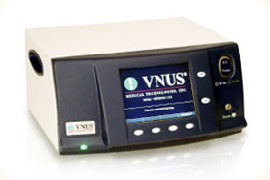 The surgeon inserts a small amount of local anaesthetic into the vein to numb the area before inserting a catheter. Once the catheter is positioned manual pressure is applied to the catheter and vein 20 seconds at a time during the treatment phase. After the procedure small steri-strips and stockings will be applied to the treated leg.
The whole process should take 40-60 minutes and patients are able to mobilise immediately. The stockings are worn day and night for 2 weeks. There are no restrictions on exercising or activity provided the stocking is worn continuously for 2 weeks.
The main equipment used for this procedure, a Radio-frequency Generator, was purchased by SSCF in April 2009 and donated to the Royal Sussex County Hospital.
Stroke
Stroke is a devastating neurological disease affecting all age groups. In Sussex approximately 3000 people will suffer a stroke each year. Stroke is caused by damage to blood vessels in the brain, either through small clots (cerebral embolism) or bleeding (cerebral haemorrhage).
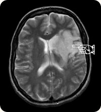
The picture on the right shows a brain scan of a patient suffering a cerebral embolism, the arrow pointing to the area of brain damage caused by the stroke. This stroke sufferer would have sustained loss of power and sensation in the limbs on the opposite side of the body to the area of brain damage. Strokes can also cause confusion, loss of speech and loss of balance.
Stroke can be prevented by attention to risk factors for vascular disease; stopping smoking, control of blood pressure and cholesterol and detection and treatment of diabetes. People who have suffered a previous stroke or mini-stroke (TIA), or are known to have vascular disease, will benefit from a daily aspirin (after discussion with their doctor). Some may benefit from an operation on the carotid arteries (after assessment by carotid ultrasound).
Those with an irregular heartbeat (atrial fibrillation) may also benefit from treatment with warfarin (after discussion of the risks and benefits of treatment with their doctor).
In the initial stages the disability caused by a stroke can appear overwhelming. However, it is important to remember that recovery can be very good, and will continue over months, and sometimes years.
Patients may be helped by hospital admission, and by assessment and treatment by stroke therapists, for example physiotherapists, occupational therapists and speech and language therapists.
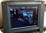
Equipment for diagnosis
Ultrasound
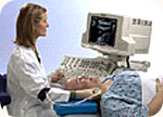 Many of the vascular laboratory tests employ ultrasound. This is a non-invasive way of examining structures within the body (ie no injection is required). Gel is placed on the skin overlying the area of interest and a probe is gently placed on the gel to examine the blood vessel below.
Doppler
A simple ultrasound probe used to listen to blood flowing in the arms and legs. A cuff around the limb records the blood pressure.
Duplex Scan
 A more complex device which gives both a picture of the vessel and information about the blood flow within that vessel. Very useful for assessing the severity of disease in both narrowed and aneurysmal vessels.
Transcranial Doppler (TCD)
Records blood flow in the head. Reveals emboli (small clots) that may come from the heart or neck arteries. Used in the operating theatre to monitor blood flow during carotid artery surgery.
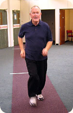
Treadmill
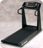 Conveyor belt similar to those seen in gyms. Records the speed and distance walked. Blood pressures in the feet may drop after exercise if the arteries in the leg are narrowed, this can be measured with Doppler.
Aneurysm Screening
Aortic aneurysms are more common in men, particularly those with a family history of the condition. The laboratory runs a screening programme to detect these in men over 65 using a quick and simple ultra sound examination. Patients with small aneurysms are followed up in the laboratory with further scans, those with larger vessels are referred for surgical assessment.
Looking after your health
- Stop smoking
- Eat a healthy diet
Avoid excess fat, particularly animal fat. Eat plenty of fresh fruit and vegetables (5 portions/day). - Exercise
Take regular exercise 3 times a week for 40 minutes at least. Walking, cycling or swimming are all recommended. - Watch your weight
Keep your weight down (especially when you stop smoking). Remember if you are taking in more calories than you need, your weight will go up. - Medication
Keep your diabetes, high blood pressure or high cholesterol under control. Take a small dose of aspirin every day if recommended by your doctor.